


- Optimized compression for highest image quality and patient comfort
- Optimized dose for high image quality and lowest possible dose
Lives can be saved by early detection of breast cancer. Mammographic screening and diagnostic technologies are now available to more women than ever before, enabling physicians to detect suspected cancers at their earliest, most treatable stages.
|
As one of the most effective methods to detect breast cancer, Mammography is used today for screening and for advanced diagnosis, including stereotactic biopsy. Recent studies show that early detection of breast cancer with Mammography can reduce mortality rates by 30%. However, breast cancer is still profound worldwide, the leading cancer in women. For Siemens, facts such as these are not just statistics; they give our work meaning. |
With Siemens' proven Mammography technology, the medical community is equipped to successfully address the increasing challenges resulting from today's disease incidence and changing economic circumstances. Quality patient care requires reliability and accuracy, balanced with patient comfort and cost-effectiveness.
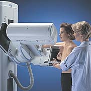
The Siemens MAMMOMAT systems are designed with superior accessibility allowing excellent patient- and breast- positioning for optimal mammography images. The attractive, patient-friendly and comfortable design allows the patient to sit, stand or lie down during the examination. The superb system design also allows easy access for the physician to perform stereotactic biopsy procedures.
When women complained about uncomfortable traditional mammography, Siemens listened. We developed Opcomp®, our exclusive, automatic compression system, designed to determine the precise amount of breast compression necessary. Based on individual breast characteristics, such as density, Opcomp produces uniform tautness and optimal image quality.
With Opcomp, the compression is increased as long as the breast is soft and pliable - then it stops. Thus, it is less likely that too little compression is used - producing inferior images - or too much compression is used, making the patient uncomfortable and anxious without improving image quality.
|
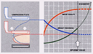
The left frame of this figure shows the breast during compression. The blue line
on the graph shows decreasing breast thickness as compression is applied. As the breast
becomes compressed, the orange line shows increasing image quality and the green line
shows increasing patient discomfort. Opcomp automatically determines optimal compression
for the best mammogram - and stops there (white arrow), sparing further discomfort.
|
|
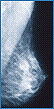
As mammography is used for both screening and diagnostic procedures, it is essential that dose exposure be kept as low as possible. This is especially important in screening programs for younger women, as their breasts normally have higher density, typically requiring higher dose. Left: medio-lateral oblique mammogram |
As part of the Siemens C.A.R.E. initiative, the MAMMOMAT 3000 Nova and Mammomat 1000 employ a number of technical features and applications designed to make mammography safer for both the patient and the operator. These include Opdose®, the automatic x-ray beam quality selection system that provides optimized X-ray parameters (selection of anode, filter, dose), tailored to individual breast characteristics.
C.A.R.E. = Combined Application to Reduce Exposure
What’s the most exciting current R&D project in Mammography? FFDM, or Full-Field Digital Mammography. Some day in the not-too-distant future, mammograms will no longer be acquired and shown on film, but rather digitally, on a computer screen, or workstation. This is not as simple as it sounds, though, as a very high-quality, high-resolution detector must replace the object table (film-holder, where the breast is positioned), the image must be transferred quickly and in full resolution to the monitor, and the detector must be ready within seconds to acquire the next image. Siemens is currently investing significant resources into Full Field Digital Mammography development, and has the first prototype installed and in clinical use.
More on MAMMOMAT 3000 Nova and Minimally Invasive Breast Biopsy (Opdima and Mammotome)
