


- How Breast MRI is Performed
- Benefits of an MRI Exam of the Breast
- Limitations to an MRI Exam of the Breast
Magnetic resonance breast imaging (MRI, MR) has been approved by the U.S. Food and Drug Administration (FDA) since 1991 for use as a supplemental tool, in addition to mammography, to help diagnose breast cancer. Breast MRI is an excellent problem-solving technology. It is often used to investigate breast concerns first detected with mammography, physical exam, or other imaging exams. MRI is also excellent at imaging the augmented breast, including both the breast implant itself and the breast tissue surrounding the implant (abnormalities or signs of breast cancer can sometimes be obscured by the implant on a mammogram). MRI is also useful for staging breast cancer, determining the most appropriate treatment, and for patient follow-up after breast cancer treatment.
In addition to its role as a diagnostic tool, researchers have been investigating whether breast MRI may be useful in screening younger women at high risk of breast cancer. Most women under 40 years of age do not require any breast imaging. However, the American Cancer Society recently recommended that women at very high risk of developing breast cancer have annual breast MRI exams in addition to annual mammograms to increase the likelihood that breast cancer will be detected early, when the chances of survival are greatest. Because MRI is more sensitive than mammography, it can help detect cancer that may be missed by mammography. However, because this increased sensitivity can also lead to false positive results, which requires breast biopsy procedures, the American Cancer Society does not recommend MRI for all women.
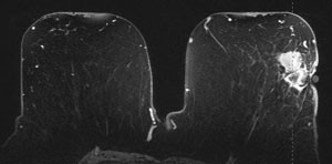
MRI image of the breast, showing a lesion. Image courtesy of Siemens Medical.
Unlike mammography which uses low dose x-rays to image the breast, MRI uses powerful magnetic fields and radio waves to create images of the breast. The MRI system is able to switch magnetic fields and radio waves to achieve views in any plane and from any orientation while x-ray mammography requires re-orientation of the breast and mammography system for each view desired.
The main component of most MRI systems is a large tube-shaped or cylindrical magnet. To begin the MRI exam, the patient is positioned on a special table inside the MRI system opening where a magnetic field is created by the magnet. Each total MRI exam is typically comprised of a series of 2 to 6 sequences, with each sequence lasting between 2 and 15 minutes. An "MRI sequence" is an acquisition of data that yields a specific image orientation and a specific type of image appearance or "contrast."

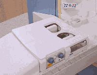
A recent advance in MRI breast imaging is the CP Breast Array Coil, which allows for bilateral breast imaging and improved differentiation between various breast tissue. The patient is placed directly on the table and the technologist has visual control of breast position through a transparent window. Images courtesy of Siemens Medical.
During the examination, a radio signal is turned on and off, and subsequently, the energy which is absorbed by different atoms in the body is echoed or reflected back out of the body. These echoes are continuously measured by the MRI scanner. A digital computer reconstructs these echoes into images of the breast. The tapping heard during the MRI exam is created when "gradient coils" are switched on and off to measure the MRI signal reflecting back out of the patient's body. A benefit of MRI is that it can easily acquire direct views of the breast in almost any orientation while mammography requires re-orientation of the breast and mammography system for each view desired. An MRI exam of the breasts typically takes between 30 and 60 minutes.
The most useful MRI technique for breast imaging uses a contrast material called Gadolinium DTPA, which is injected into a vein in the arm before or during the exam to improve the quality of the images. This contrast agent helps produce stronger and clearer images and "highlight" any abnormalities.
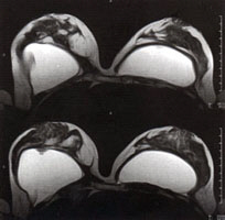
Transverse high-resolution MRI scan of breast and implants. Note the implant twisting on the upper (left) image and the implant valve on the lower (left) image.
