


- What is Vacuum-Assisted Biopsy?
- How is Vacuum-Assisted Biopsy Performed?
- How Should Patients Prepare for Vacuum-Assisted Biopsy?
- What Should Patients Expect After Vacuum-Assisted Biopsy?
- What are the Advantages and Disadvantages to Vacuum-Assisted Biopsy?
The relatively new vacuum-assisted breast biopsy is a percutaneous ("through the skin") procedure that relies on stereotactic mammography or ultrasound imaging. Stereotactic mammography uses computers to pinpoint the exact location of a breast mass based on mammograms (x-rays) taken from two different angles. The computer coordinates will help the physician to guide the needle to the correct area in the breast. With ultrasound, the radiologist or surgeon will watch the needle on the ultrasound monitor to help guide it to the area of concern. The patient will either by positioned in the upright or prone (face down) position for a vacuum-assisted biopsy.
Vacuum-assisted biopsy is a minimally invasive procedure that allows for the removal of multiple tissue samples. However, unlikecore needle biopsy, which involves several separate needle insertions to acquire multiple samples, the special biopsy probe used during vacuum-assisted biopsy is inserted only once into the breast through a small skin nick made in the skin of the patient's breast.
 |
The Mammotome Biopsy System made by Johnson & Johnson Ethicon Endo-Surgery |
Two companies currently manufacturer vacuum-assisted breast biopsy systems, and often, vacuum-assisted biopsy will be referred to by the brand name: either Mammotome made by Johnson & Johnson Ethicon Endo-Surgery or MIBB (which stands for Minimally Invasive Breast Biopsy) made by Tyco/United States Surgical Corporation. In 1999, a hand-held version of the Mammotome was also approved by the U.S. Food and Drug Administration (FDA).
First, the skin of the breast is cleaned. Then, a small amount of local anesthetic (lidocaine), similar to what one might have at a dentist’s office, is injected into the skin and deeper tissues of the breast using a small hypodermic needle. Under stereotactic or ultrasound guidance, the radiologist or breast surgeon positions the special breast probe into the area of the breast where the lesion (abnormality) is located.
After the probe has been properly positioned, a vacuum line draws the breast tissue through the aperture of the probe into the sampling chamber of the device. Once the tissue is in the sampling chamber, the rotating cutting device is advanced and a tissue sample is captured. The tissue sample is then carried through the probe to the tissue collection area (a standard pathology tissue cassette).
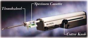
After a tissue sample is captured, the radiologist or surgeon then rotates the thumbwheel of the probe, moving the sampling chamber approximately 30 degrees to new position. The entire cycle is repeated, until all desired areas have been sampled (typically, eight to 10 samples of breast tissue are taken 360 degrees around the lesion).
 |
| Mammotome Hand-Held Biopsy System, Courtesy of Johnson & Johnson Ethicon Endo-Surgery |
When a sufficient number of tissue samples have been collected, the radiologist or surgeon
will remove the probe and apply pressure to the biopsy site. An adhesive bandage will be
applied to the skin nick. In some cases, a small sterile clip will be placed into the
biopsy site of the breast to mark the location in case a future biopsy is needed. This
microclip is left inside the breast and causes no pain, disfigurement, or harm to the
patient. After the biopsy is complete, the tissue samples will be sent to the pathology
laboratory for diagnosis.
